Physiology of the eye
Retina
The retina is a thin layer at the backside of the eye comprised of photoreceptors and cells for signal processing.
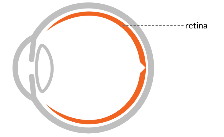
Parts of the retina
Cones
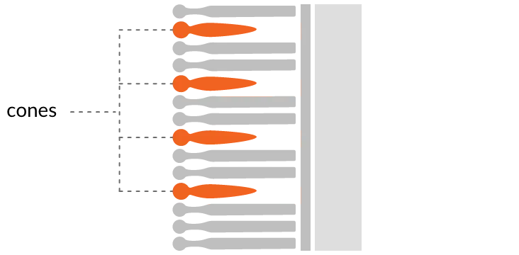
Cones have photopigments that are sensitive to three distinct wavelengths of light. The retina is comprised of approximately 7 million cones.
When only cones are functioning (sunlight) it is called photopic vision. When both cones and rods are functioning (twilight), it is called mesopic vision. People with color vision deficiencies may have missing or abnormally functioning cones.
Rods
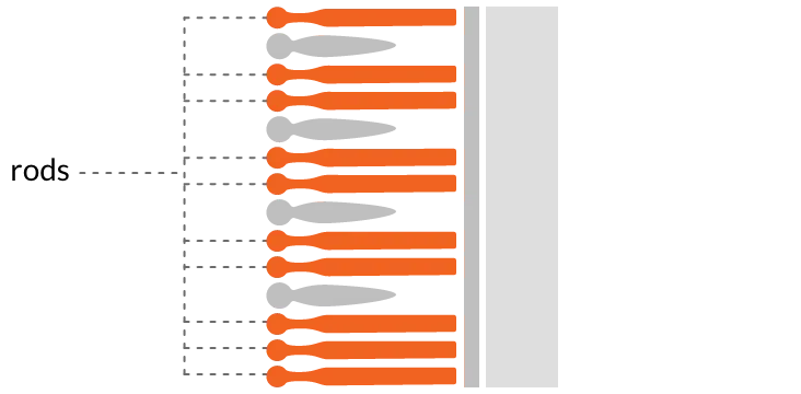
Rods are a type of photoreceptor that works at lower levels of light. The retina is comprised of approximately 120 million rods.
When only the rods are functioning, it is called scotopic vision. Rod-only vision is monochromatic and can be attributed to color vision deficiencies.
Fovea
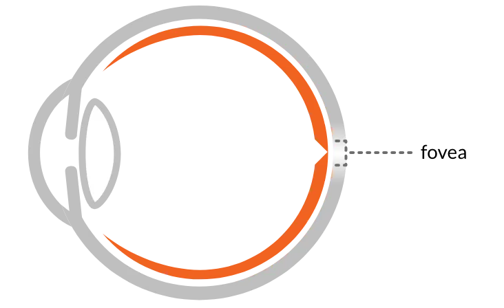
The fovea is the area of the retina that has the best visual acuity and color vision. The fovea has the highest concentration of cones and the lowest concentration of rods.
The fovea is protected from intense exposure to short-wavelength energy by a yellow filter called the macula.
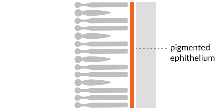
Pigmented epithelium
The pigmented epithelium is the bottom layer of the retina, and is pigmented in order to absorb any remaining light that was not processed by the cones and rods.
Sources:
- Mark D. Fairchild (2013). Color Appearance Models. 3rd edition. John Wiley & Sons, Ltd. Pages 5-13
- R.W.G. Hunt, M.R. Pointer (2011). Measuring Colour. John Wiley & Sons, Inc. Pages 448
- How eyes work. . https://www.nei.nih.gov/learn-about-eye-health/healthy-vision/how-eyes-work